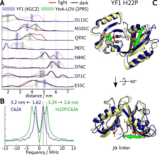
Structure of YF1. a Structure of YF1 in its dark-adapted state as
4.6 (734) · $ 6.50 · In stock

Download scientific diagram | Structure of YF1. a Structure of YF1 in its dark-adapted state as resolved by X-ray crystallography 13. The location of the different domains, of the flavin mononucleotide (FMN), of the cofactor adenosine diphosphate (ADP), and of the phosphoaccepting histidine 161 are indicated. b Light induced conformational changes of the LOV photosensor domain refined from X-ray solution scattering 22. The changes are maximal at the C-termini that feed into the Jα helices (dashed arrows). The coloring is according to the root mean square deviation of the alpha carbons from publication: Sequential conformational transitions and α-helical supercoiling regulate a sensor histidine kinase | Sensor histidine kinases are central to sensing in bacteria and in plants. They usually contain sensor, linker, and kinase modules and the structure of many of these components is known. However, it is unclear how the kinase module is structurally regulated. Here, we use | Secondary Protein Structure, Bacterial Proteins and Protein Conformation | ResearchGate, the professional network for scientists.
Blue-light reception through quaternary transitions
Thermodynamics of photoreceptors. (A) Absorption of light drives the

Signal transduction in light–oxygen–voltage receptors lacking the adduct-forming cysteine residue

Oskar BERNTSSON, PostDoc Position, Doctor of Philosophy, MAX IV Laboratory, Lund
Toxins, Free Full-Text

Internal force minimization-based adaptive beam string structure

Ontogeny of the Postorbital Region in Tarsiers and Other Primates - DeLeon - 2016 - The Anatomical Record - Wiley Online Library

Gemma NEWBY, Application Scientist, PhD Chemistry, Xenocs, Sassenage, Science and Application

A) Structure of YF1 with a LOV photosensor dimer, consisting of A

Biomolecules, Free Full-Text

Light-Oxygen-Voltage (LOV)-sensing Domains: Activation Mechanism and Optogenetic Stimulation - ScienceDirect