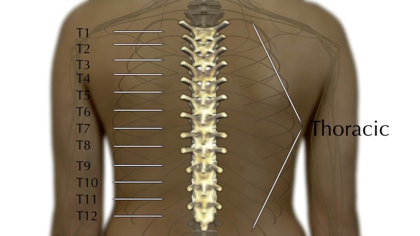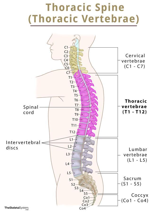
A thoracic spine MRI scan examines the middle section of your spine between the neck and lower back.

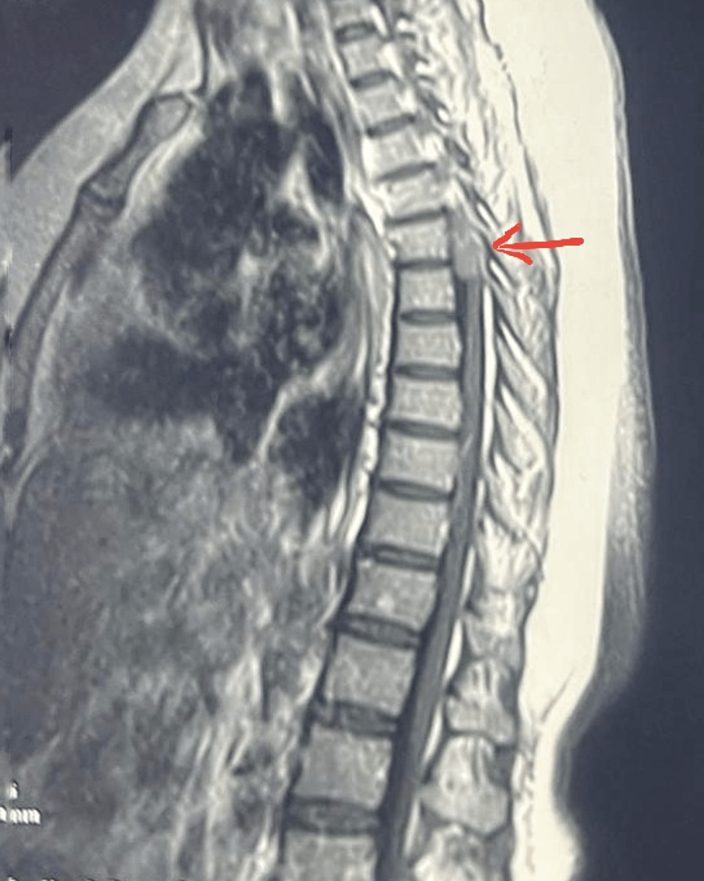
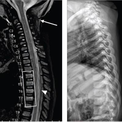
MRI OF THORACIC SPINE HISTORY A 53-year-old woman, presented with paresis and paresthsia of both legs. Longitudinally extensive T2W hyperintensity at spinal cord . 15763120 Stock Photo at Vecteezy
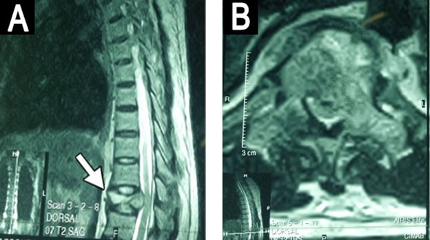
Cureus, Intramedullary Thoracic Spinal Cord Abscess Mimicking an Intramedullary Tumor: A Case Report
Whole-spine MRI uncovers abusive head trauma injuries in children
Surgical Neurology International

Close Mri Thoracic Spine 2view Showing Stock Photo 1057683110

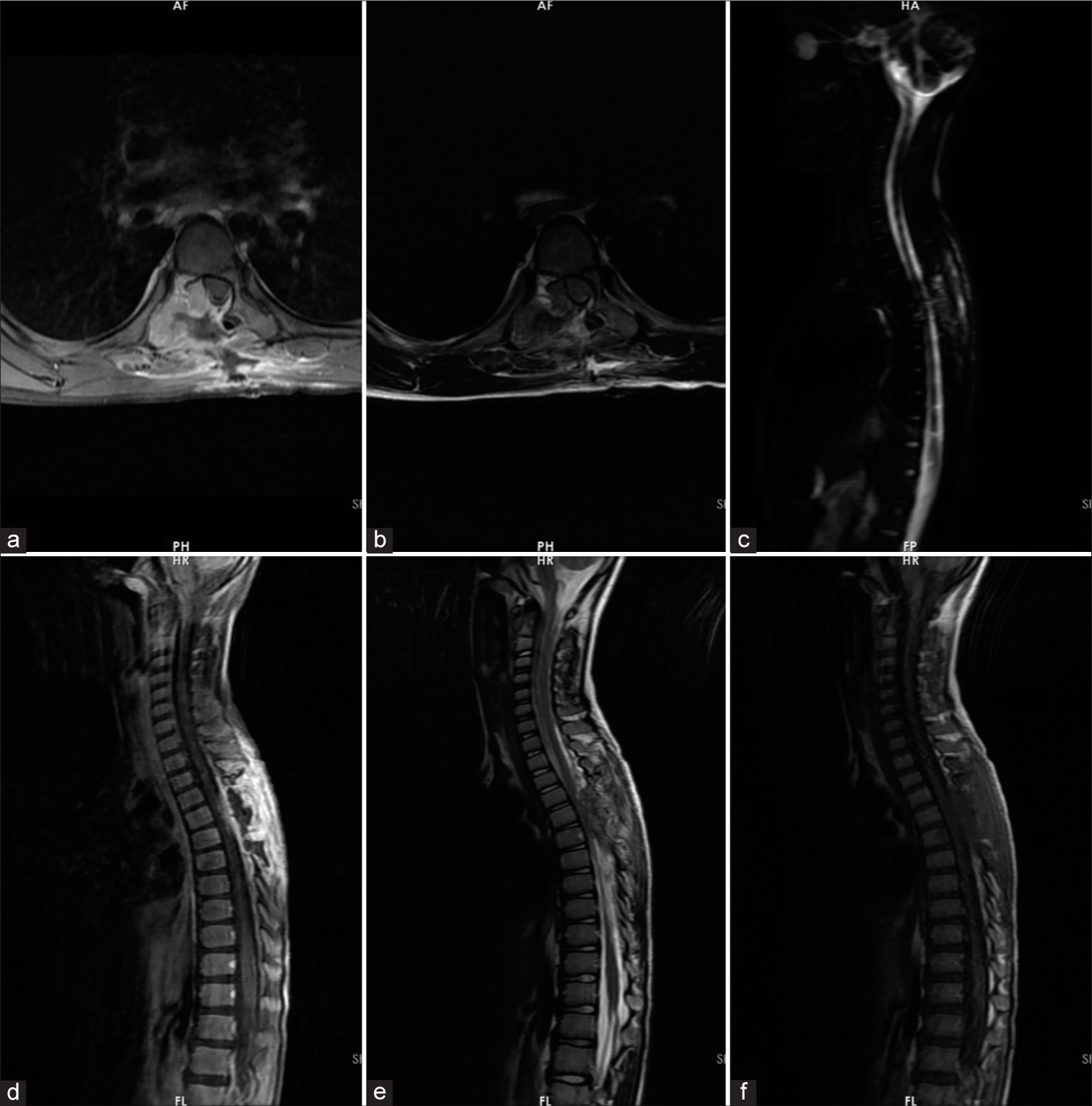
MRI features of spinal chronic recurrent multifocal osteomyelitis/chronic non-bacterial osteomyelitis in children
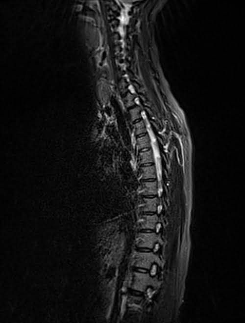
MRI thoracic spine STIR sagittal images
AJNS – African Journal of Neurological Sciences » SURGICAL MANAGEMENT OF PRIMARY OSSEOUS THORACIC SPINE HEMANGIOPERICYTOMA IN A LOW RESOURCE SETTING: A CASE REPORT.

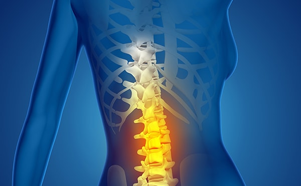
Thoracic Herniated Disc

MRI scan of thoracic (upper row) and lumbar spine (lower row) with and