Figure 6 from Femoral Hernia: A Review of the Clinical Anatomy and
4.7 (660) · $ 26.50 · In stock

Figure 6. Femoral hernia repair in clean operation. (a) The narrow side of the mesh is sutured to Cooper’s ligament; (b) The mesh is sutured to the iliopubic tract or shelving portion of the inguinal ligament; (c) The posterior wall of the inguinal canal is reinforced, as in Lichtenstein’s repair. - "Femoral Hernia: A Review of the Clinical Anatomy and Surgical Treatment"
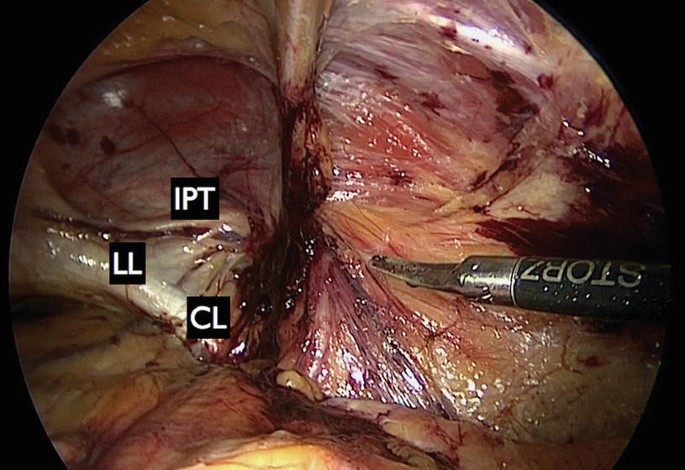
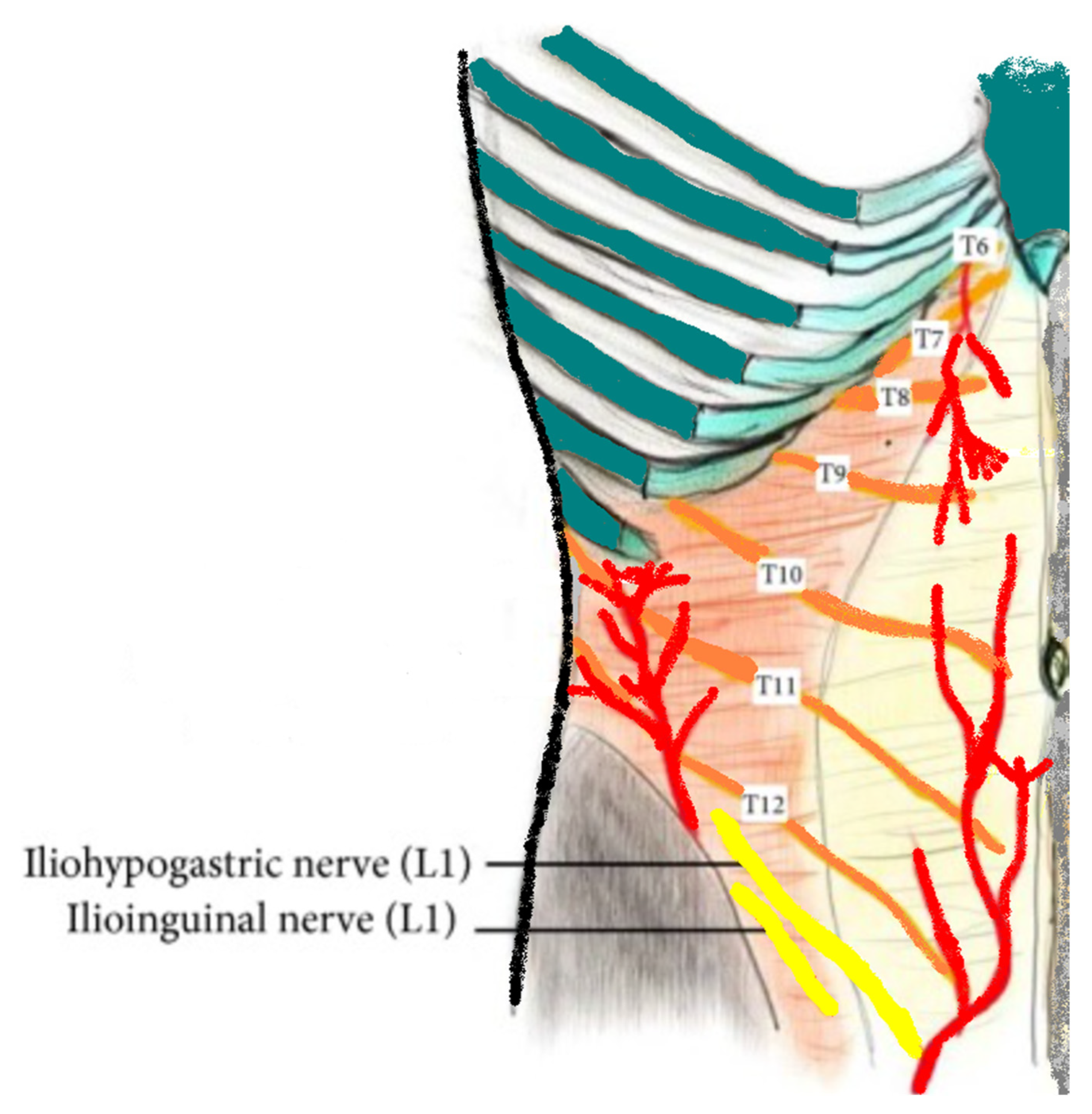
8 Anatomical basis of the myopectineal orifice (Fruchaud) or inner

Femoral Hernia - A Review of Clinical Anatomy

Femoral Hernia: A Review of the Clinical Anatomy and Surgical Treatment

A Endoscopic view of a left femoral hernia (o) in a female patient

Femoral Hernia - A Review of Clinical Anatomy

d3i71xaburhd42.cloudfront.net/7f672b1a5e914d2febb0

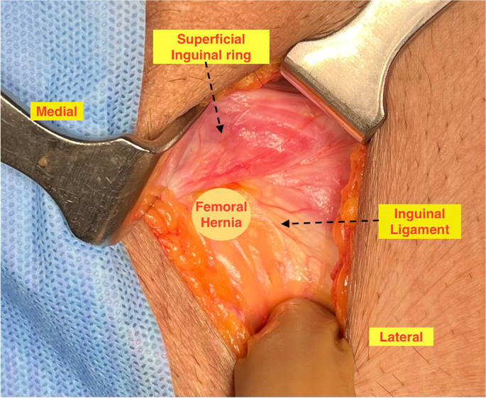
Presence of the appedix vermiformis in the femoral hernia.
Surgical Techniques Development, Free Full-Text

Intraperitoneal inspection. *: The inguinal ligament; L: Left; R
Femoral Hernia - A Review of Clinical Anatomy

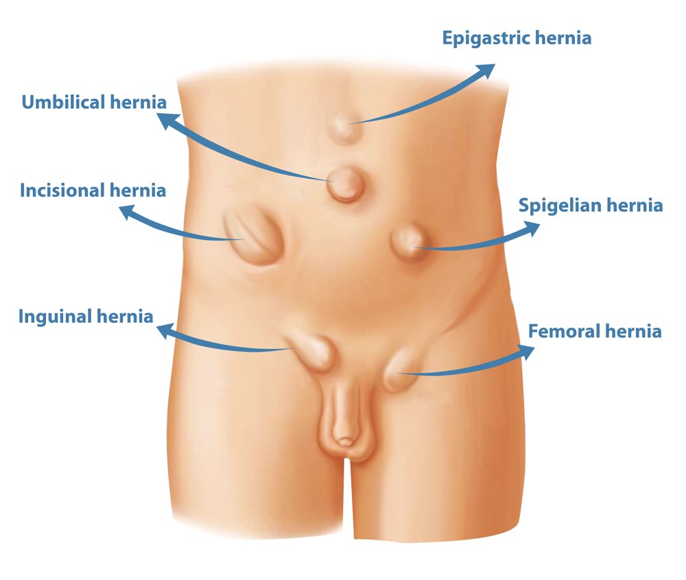
Figure, Abdominal Hernias Contributed by T Silappathikaram] - StatPearls - NCBI Bookshelf
Hernia - Physiopedia

Figure 1 from Femoral Hernia: A Review of the Clinical Anatomy and Surgical Treatment
Inguinal ligament: Attachments, function and relations

AIS Channel · Hernia Surgery