Classification of femoral hernia. adventitia is ligament into
4.7 (536) · $ 7.99 · In stock

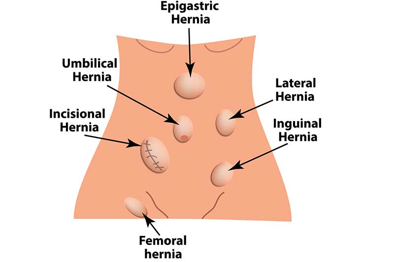
Download scientific diagram | Classification of femoral hernia. adventitia is ligament into continuing fascia. superiorlly, the ligament 3 2 1 T . . . funnel he M M L psoas three ateral edial iddle boundaries I n peritoneal the parts: and and of part shape part part transverse lacunar the area pectineal femoral is with with femoral femoral of of that lining femoral femoral the the ligament fascia, is vein thigh femoral muscle, vessels. canal. of formed vein, the artery, posterior laterally. about medially, femoral canal F and from emoral 3 cm the to are T canal caudally the the below he inguinal sides region fascia femoral anteromedial fuses the of is pectineal ligament the covering inguinal with divided canal iliac the from publication: Tension free femoral hernia repair with plug | Objective To investigate the conventional technique involves treatment of femoral hernia an approximation inguinal ligament to pectineal ligament. In technique which uses mesh closure for femoral canal without tissue tension.MethodsA prospective study from January 01. | Femoral Hernia, Repair and Mesh | ResearchGate, the professional network for scientists.

Femoral hernia - Clinical Anatomy & Operative Surgery
Femoral Hernia
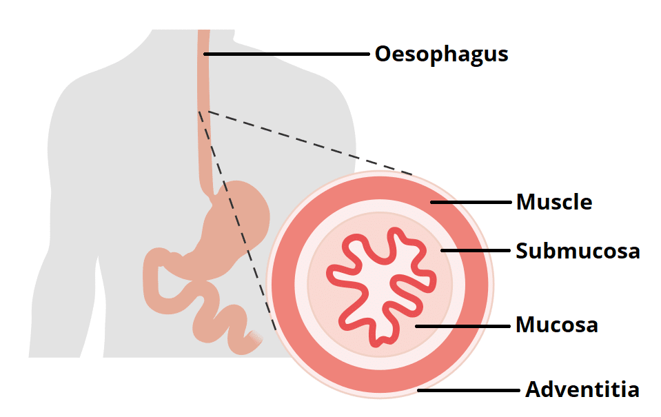
The Oesophagus - Location - Sphincters - TeachMeAnatomy

Milivoje VUKOVIĆ, General and Digestive Surgeon

The formation of Prolene mesh plug.

Classification of femoral hernia. adventitia is ligament into

Figure 4 from Femoral Hernia: A Review of the Clinical Anatomy and Surgical Treatment

JaypeeDigital

Inguinal and femoral hernias (mechanism of disease)

Femoral hernia, Radiology Reference Article

PDF) Tension free femoral hernia repair with plug

Surgical Anatomy of Groin and Groin Hernia