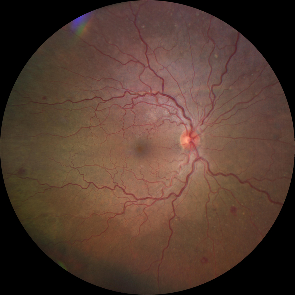
Ultra-wide-field fundus photographs and ultra-wide-field
4.8 (763) · $ 10.00 · In stock

Download scientific diagram | Ultra-wide-field fundus photographs and ultra-wide-field fluorescein angiographic imaging of ocular toxocariasis. (A) A granuloma with mild vitreous opacity. (B) A tractional retinal fold with localized tractional retinal detachment. (C) Diffuse peripheral vascular leakage. (D) A prominent optic disc leakage. from publication: The Clinical Characteristics of Ocular Toxocariasis in Jeju Island Using Ultra-wide-field Fundus Photography | Toxocariasis, Ocular and Photography | ResearchGate, the professional network for scientists.

Deep learning can generate traditional retinal fundus photographs using ultra-widefield images via generative adversarial networks - ScienceDirect
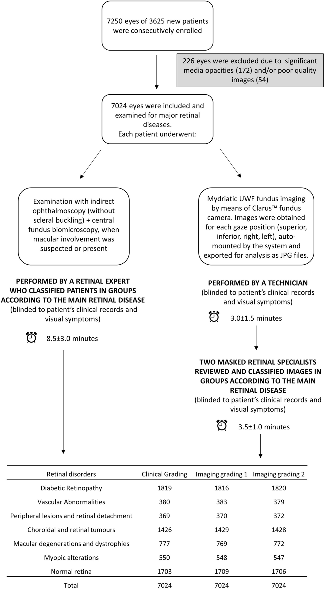
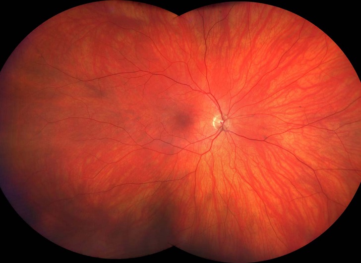
Ultra-wide-field fundus photography compared to ophthalmoscopy in diagnosing and classifying major retinal diseases

Sang-Yoon Lee's research works Gachon University, Seongnam-si (kyungwon) and other places

Eun Kyoung Lee's research works Dongguk University, Seoul and other places

PDF) The Clinical Characteristics of Ocular Toxocariasis in Jeju Island Using Ultra-wide-field Fundus Photography
ZEISS CLARUS 500 Fundus Camera

Ultra-wide-field fundus images with overlay of the Early Treatment
What Is Ultrawide-Field Imaging Really Showing Us?
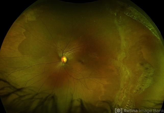
Ultra-Wide Field Fundus Photography Showing Lattice Degeneration - Retina Image Bank
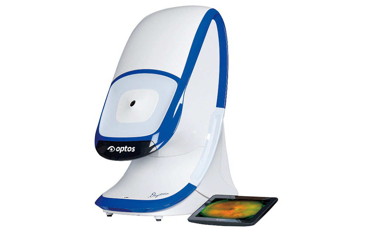
Ultra-Wide Field Retinal Imaging Device, Product Technology

Comparison of true-colour wide-field confocal scanner imaging with standard fundus photography for diabetic retinopathy screening

Ultra-wide field pseudocolor fundus image (after stereographic

Sang-Yoon Lee's research works Gachon University, Seongnam-si (kyungwon) and other places

Ultrawide-field color fundus photography (1) and ultrawide-field fundus

Eun Kyoung Lee's research works Dongguk University, Seoul and other places