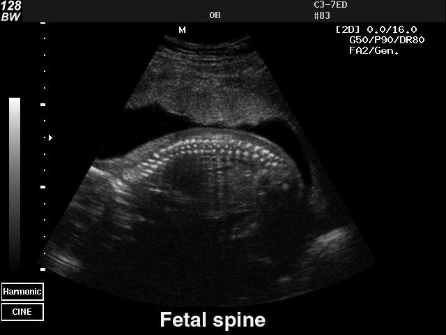
Grey scale imaging (ultrasound), Radiology Reference Article
4.7 (555) · $ 17.00 · In stock

Commonly referred to as B (brightness) mode, the use of grey scale imaging in ultrasound renders a two-dimensional image in which the organs and tissues of interest are depicted as points of v
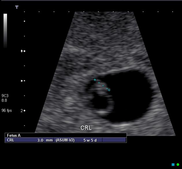
Crown rump length, Radiology Reference Article

Posterior Fossa Horns in Hurler Syndrome: Prevalence and Regression

Multi-contrast submillimetric 3 Tesla hippocampal subfield segmentation protocol and dataset

ULTRASOUND CORNER: RANGE AMBIGUITY ARTIFACT - O'Brien - 2001 - Veterinary Radiology & Ultrasound - Wiley Online Library

SciELO - Brasil - Proposal for computer-aided diagnosis based on ultrasound images of the kidney: is it possible to compare shades of gray among such images? Proposal for computer-aided diagnosis based on

Grayscale ultrasonographic image demonstrating normal kidney size and
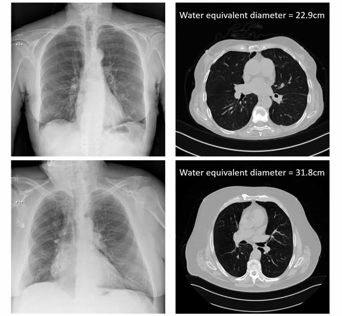
Estimating patient size from X-ray data improves radiation risk assessment – Physics World

Grayscale Ultrasound Artifacts

PDF] From Grey Scale B-Mode to Elastosonography: Multimodal Ultrasound Imaging in Meningioma Surgery—Pictorial Essay and Literature Review
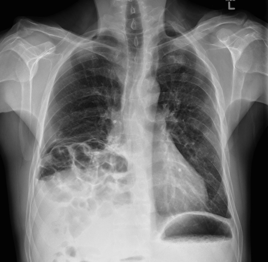
Diaphragmatic hernia - Radiology at St. Vincent's University Hospital
Gray-scale features of markedl [IMAGE]
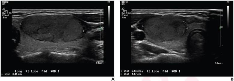
TI-RADS, Algorithm Guide Diagnoses of Pediatric Thyroid Nodules on Ultrasound

Grayscale Ultrasound Artifacts
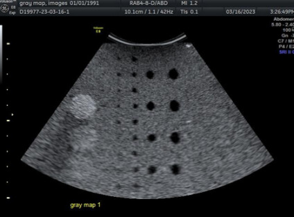
50 More Shades of Gray: About Gray Maps - Ultra Select Medical
On the superior panel, 2D gray scale sonography demonstrates a